Wednesday, December 23, 2015

The Physiologically Difficult Airway

Jarrod M. Mosier, MD , Raj Joshi, MD , Cameron Hypes, MD, Garrett Pacheco MD, Terrence Valenzuela, MD, John Sakles MD.
                                      University of Arizona ICU and ED Departments.
                                   Published, Western Journal of Emergency Medicine 2015
Full text available via open access http: escholarship.org/uc/uciem_westjem 
DOI 10.5811/westjem/ 2015
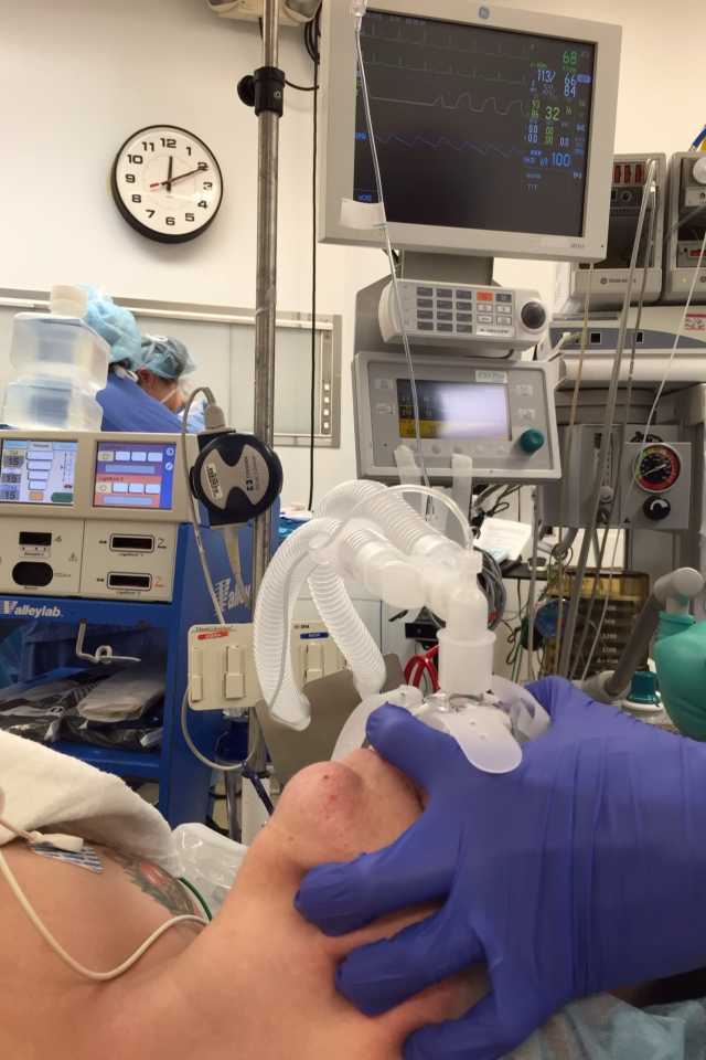
With recognition that many tools ( DL, VL, FOB, Surgical neck access),  are now available for the placement of endotracheal tubes and that CPAP , BIPAP also make a powerful contribution to oxygenation and ventilation ,  there remains another dimension of the airway management problem that needs to be addressed. 

Physiological Factors:
This important contribution to the teaching of Airway strategy underlines the four physiological states that add a complexity and risk to the Difficult Airway patient management. The special problems in the ICU and the ED are often coloured by the complex physiology of people who are suffering from profound general disorders. It is therefore fitting that this new look at the difficult Airway should come from Mosier (ICU) and Sakles (ED). Separation of these factors for special education and acute care consideration will surely make care safer in critical care areas.

1. Hypoxemia - with a patient at an unfavourable point on the oxygen dissociation curve leaving reduced margin for rapid deterioration. The pre oxygenation process becomes important prior to attempts at intubation. The use of Nasal approaches to provision of procedural oxygen are currently attracting more attention and study. These include the Thrive Hi Flo Nasal oxygen strategy, the simple use of nasal prongs (less effective but still added value) and nasal TSE PAP which uses the nose as a conduit for CPAP with a modified #2 Childrens mask.

    2. Hypotension- addressed  with standard volume optimization support  and pressor use as indicated. 

    3. Severe Metabolic acidosis - treated with disease specific therapy (i.e. Diabetic Keto-acidosis) and or other cause specific therapy such as septic state therapy.

    4. Right Ventricular failure - firstly awareness of the diagnosis is key followed by excellent strategies defined by truly expert care. The following are considered to be of value by Mosier and his team 1. Available bedside cardiac echo to assess right heart reserve allowing fluid use, 2. pre oxygenation (see above) 3. consider etomidate induction, 4. consider Norepinephrine to increase systemic pressure, and low mean airway pressure ventilation. To obtain a discussion of these outline points consult the original detail embodied in the paper itself.

Abstract


Monday, December 21, 2015

Video Laryngoscopy Improves Odds of First Attempt Success at Intubation in the ICU: A Propensity-Matched Analysis

Cameron D HypesUwe StolzJohn C SaklesRaj R JoshiBhupinder NattJosh MaloJohn W Bloom, and Jarrod M Mosier

Jarrod Mosier and John Sakles continue to be among leaders in clinical evaluation of airway management in ICU and ED areas. This article reinforces the growing understanding that in ICU , where most caregivers are no longer from an anesthesiology training base but from various other disciplines, the Video Laryngoscope is easier to learn and use. The skill retention is also superior using VL so overall performance for first pass success is superior to DL.
The Abstract for ICU application is as follows:

Abstract

 Rationale: Urgent tracheal intubation is performed frequently in intensive care units and incurs higher risk than when intubation is performed under more controlled circumstances. Video laryngoscopy may improve the chances of successful tracheal intubation on the first attempt; however existing comparative data on outcomes are limited. 
Objectives: To compare first attempt success and complication rates during intubation when using video laryngoscopy compared to traditional direct laryngoscopy in a tertiary academic medical intensive care unit. Methods: We prospectively collected and analyzed data from a continuous quality improvement database of all intubations in one medical intensive care unit between January 1st, 2012 and December 31st 2014. Propensity-matching and multivariable logistic regression were used to reduce the risk of bias and control for confounding.

 Measurements and Main Results: A total of 885 intubations took place over the study period. Of these, 673 (83.2%) were performed using video laryngoscopy and 
136 (16.8%) using direct laryngoscopy. 
First attempt success with video laryngoscopy was 80.4% (95% CI 77.2 to 83.3%) compared and 65.4% (95% CI: 56.8 to 73.4%) for intubations performed with direct laryngoscopy, p=<0.001. In a propensity-matched analysis, the odds ratio for first attempt success with video laryngoscopy vs. direct laryngoscopy was 2.81 (95% CI 2.27 to 3.59). The rate of arterial oxygen saturation events during the first intubation attempt was significantly lower for video laryngoscopy than for direct laryngoscopy (18.3% vs. 25.9%, p=0.04). The rate of esophageal intubation during any attempt was also significantly lower for video laryngoscopy (2.1% vs. 6.6%, p=0.008). 
Conclusions: Video laryngoscopy was associated with significantly improved odds of first attempt success at tracheal intubation by non-anesthesiologists in a medical intensive care unit. Esophageal intubation and oxygen desaturation complicating intubation occurred less frequently with the use of video laryngoscopy. Randomized clinical trials are needed to confirm these findings.


Read More: http://www.atsjournals.org/doi/abs/10.1513/AnnalsATS.201508-505OC#.VniHCTa0GfM   


Tuesday, December 1, 2015

Ventrain- A novel approach to ventilation through small bore cannulas by Professor Dietmar Enk

The Ventrain device is designed to take advantage of the Bernoulli Principle to 
create a control that will use the pressure of an Oxygen source to create 
positive pressure inflow and when the appropriate orifice is blocked a negative pressure for expiration is created by the passage of air over the entrance to the endotracheal tube. This is explained in the Text.

This substantially improves the potential to ventilate through small caliber cannulas. 

The simplicity and brilliance of the design suggests an important role that will improve difficult airway management and also possibly a number of other situations.

Sunday, November 29, 2015

Comparison of the GlideScope Highly Angled Video Laryngoscope with the Storz D- Blade for first pass success in patients with known predictors of difficult airway randomized .


Michael Aziz MD , Ron Abrons MD, Davide Cattano MD, Emine Bayman PhD, David Swanson MD, Carin Hagberg MD , Michael Todd MD, and Ansgar Bambrink MD PhD.

This study is done with a precision that is an example of the best work in sorting out the variables in a complex group of patients treated in excellent hospitals. I commend this paper as essential reading to all airway managers  without comment as the work speaks for itself.

Saturday, November 28, 2015

Macewen Medal to Dr John Allen Pacey MD FRCSc at World Airway Management Meeting , Dublin, Nov. 2015


Acknowledgements

This medal represents the combined efforts of a great many people, while I cannot mention each great contribution the following are significant.

The teams of Saturn Biomedical Systems and our distributors such as Vitaid Company led by Will Stewart.
Awni Ayoubi who managed the roll out  of a manufacturable GlideScope and co-creation of the Saturn Biomedical Systems Company.
The Great teams at Verathon Medical both in Canada where creation and manufacture of 33+ blades occurred and in Bothell Washington where the GlideScope was taken to the world.

The great contributions of all of our Medical Experts including Dr. Richard Cooper, Dr. John Doyle, and Dr. Mike Bishop who produced  pivotal studies and documented the value of the device.
The Society for Airway Management was very important in identifying the role of GlideScope highly angled video Laryngoscope and helped spread the technology to their peers since 2001.
DAS in UK and ESA which included the Glidescope in so many workshops since 2003 that they remain uncounted.
Finally my wife Doris, an ICU and PACU nurse, who was a willing subject for many preclinical application tests . Without Doris support, there would be no prize contribution.

Saturday, September 19, 2015

Glidescope Video Laryngoscope Training Series - #3

Griesdale makes a significant point in this paper with the
Can J Anaesth. 2012 Jan;59(1):41-52. 
Glidescope® video-laryngoscopy versus direct laryngoscopy for endotracheal intubation: a systematic review and meta-analysis.


Abstract
INTRODUCTION:

The Glidescope(®) video-laryngoscopy appears to provide better glottic visualization than direct laryngoscopy. However, it remains unclear if it translates into increased success with intubation.
METHODS:
We systematically searched electronic databases, conference abstracts, and article references. We included trials in humans comparing Glidescope(®) video-laryngoscopy to direct laryngoscopy regarding the glottic view, successful first-attempt intubation, and time to intubation. We generated pooled risk ratios or weighted mean differences across studies. Meta-regression was used to explore heterogeneity based on operator expertise and intubation difficulty.
RESULTS:
We included 17 trials with a total of 1,998 patients. The pooled relative risk (RR) of grade 1 laryngoscopy (vs ≥ grade 2) for the Glidescope(®) was 2.0 [95% confidence interval (CI) 1.5 to 2.5]. Significant heterogeneity was partially explained by intubation difficulty using meta-regression analysis (P = 0.003). The pooled RR for nondifficult intubations of grade 1 laryngoscopy (vs ≥ grade 2) was 1.5 (95% CI 1.2 to 1.9), and for difficult intubations it was 3.5 (95% CI 2.3 to 5.5). There was no difference between the Glidescope(®) and the direct laryngoscope regarding successful first-attempt intubation or time to intubation, although there was significant heterogeneity in both of these outcomes. In the two studies examining nonexperts, successful first-attempt intubation (RR 1.8, 95% CI 1.4 to 2.4) and time to intubation (weighted mean difference -43 sec, 95% CI -72 to -14 sec) were improved using the Glidescope(®). These benefits were not seen with experts.
CONCLUSION:

Compared to direct laryngoscopy, Glidescope(®) video-laryngoscopy is associated with improved glottic visualization, particularly in patients with potential or simulated difficult airways.

Glidescope Video Laryngoscope Training Series #2 - ER Resident Training

Learning Curves for Direct Laryngoscopy and GlideScope®
Video Laryngoscopy in an Emergency Medicine Residency

John C Sakles, MD*Jarrod Mosier, MD*Asad E. Patanwala, PharmD† John Dicken, BS‡
Supervising Section Editor: John Ashurst, DO


Introduction: Our objective is to evaluate the resident learning curves for direct laryngoscopy
(DL) and GlideScope® video laryngoscopy (GVL) over the course of an emergency medicine (EM) residency training program.

Methods: 
    This was an analysis of intubations performed in the emergency department (ED) by EM residents over a seven-year period from July 1, 2007 to June 30, 2014 at an academic ED with 70,000 annual visits. After EM residents perform an intubation in the ED they complete a continuous quality improvement (CQI) form. Data collected includes patient demographics, operator post- graduate year (PGY), difficult airway characteristics (DACs), method of intubation, device used for intubation and outcome of each attempt. We included in this analysis only adult intubations performed by EM residents using a DL or a standard reusable GVL. The primary outcome was first pass success, defined as a successful intubation with a single laryngoscope insertion. 
     First pass success was evaluated for each PGY of training for DL and GVL. Logistic mixed-effects models were constructed for each device to determine the effect of PGY level on first pass success, after adjusting for important confounders.

Results: 
     Over the seven-year period, the DL was used as the initial device on 1,035 patients and
the GVL was used as the initial device on 578 patients by EM residents. When using the DL the first past success of PGY-1 residents was 69.9% (160/229; 95% CI 63.5%-75.7%), of PGY-2 residents was 71.7% (274/382; 95% CI 66.9%-76.2%), and of PGY-3 residents was 72.9% (309/424; 95% CI 68.4%-77.1%). When using the GVL the first pass success of PGY-1 residents was 74.4% (87/117; 95% CI 65.5%-82.0%), of PGY-2 residents was 83.6% (194/232; 95% CI 76.7%-87.7%), and of PGY-3 residents was 90.0% (206/229; 95% CI 85.3%-93.5%). In the mixed-effects model for DL, first pass success for PGY-2 and PGY-3 residents did not improve compared to PGY-1 residents (PGY-2 aOR 1.3, 95% CI 0.9-1.9; p-value 0.236) (PGY-3 aOR 1.5, 95% CI 1.0-2.2, p-value 0.067).
However, in the model for GVL, first pass success for PGY-2 and PGY-3 residents improved
compared to PGY-1 residents (PGY-2 aOR 2.1, 95% CI 1.1-3.8, p-value 0.021) (PGY-3 aOR 4.1, 95% CI 2.1-8.0, p<0.001).

Conclusion: 
Over the course of residency training there was no signifcant improvement in EM resident first pass success with the DL, but substantial improvement with the GVL. 

Full text available through open access at http://escholarship.org/uc/uciem_westjem
DOI: 10.5811/westjem.2014.9.23691    [West J Emerg Med 1-8].
University of Arizona, Department of Emergency Medicine, Tucson, Arizona
University of Arizona College of Pharmacy, Department of Pharmacy Practice and
Science, Tucson, Arizona
University of Arizona College of Medicine, Tucson, Arizona

Glidescope Video Laryngoscope Training Series - # 1 The need for Specific training

The training conundrum - VL and FOB

The same problem that afflicts training programs for FOB use is also present for training of VL. There is an increment of skill for both device categories that demands specific skill training and importantly competence testing. Teachers have variable approaches to teaching these devices  which colors the outcomes.
John Fiadjoe's group has produced an excellent paper showing the outcomes with the Glidescope Cobalt Paediatric VL when compared with the Flexible Scope category. The manikin study has relevance because it focusses on the operator / device interaction with a standardized Pierre Robin test base. The real patient has other confounding issues like secretions and blood that I submit could influence the equation in favor of VL but this is not the thrust of the paper.

 2015 Aug;25(8):801-6. doi: 10.1111/pan.12668. Epub 2015 Apr 27.

A randomized multi-institutional crossover comparison of the GlideScope® Cobalt Video laryngoscope to the flexible fiberoptic bronchoscope in a Pierre Robin manikin.

Abstract

BACKGROUND: 

The GlideScope Cobalt Video laryngoscope is being used more often in children with challenging laryngoscopy. There are, however, no pediatric trials comparing it to flexible fiberoptic bronchoscopy, the current accepted gold standard. This preliminary manikin study compares the first-attempt intubation success of the GlideScope Cobalt video laryngoscope to the flexible fiberoptic bronchoscope when performed by attending pediatric anesthesiologists at two major pediatric centers.

METHODS: 

This prospective randomized, crossover study evaluated 120 attempts (60 with each study device) to intubate the AirSim Pierre Robin manikin (PRM) with fiberoptic bronchoscopy and video laryngoscopy (VL). Attending pediatric anesthesiologists from two quaternary pediatric centers were eligible to participate. Each attending anesthesiologist randomly performed a single tracheal intubation attempt with one of the study devices followed by the alternate method. The primary outcome was the first-attempt success rate of tracheal intubation. Blinding was not feasible. We hypothesized that first-attempt success would be higher with fiberoptic bronchoscopy.

RESULTS: 

Thirty anesthesiologists from each center were randomized to use one of the study devices followed by the alternate method. We analyzed all participants' data. There was no overall difference in first-attempt success between VL and fiberoptic bronchoscopy (88.3% vs 85% respectively, P = 0.59). There were significant institutional differences in first-attempt success using VL (76.7% vs 100%).

CONCLUSIONS: 

There was no difference in first-attempt success of tracheal intubation using VL vs fiberoptic bronchoscopy when performed by attending anesthesiologists at two large pediatric centers. However, institutional differences exist in success rates with VL across the two centers. Results from single-center device evaluations should be verified by multi-center evaluations. A significant proportion of attending anesthesiologists lack experience with advanced airway devices; targeted education may enhance intubation success and patient safety.

Thursday, August 6, 2015

The Age of Nasal Oxygen Delivery is upon us.

The Age Of Nasal Oxygen

Goal:
 To explore the nasal Oxygen bridging methods to GlideScope intubation.
 Recent developments highlight the safety of nasal O2 strategies in clinical airway management.

The Classic Methods of Nasal Oxygen delivery are:

1. Standard nasal cannula - delivers 20-40 % O2 to Patients breathing via the nose.
2. Reservoir cannulas - more efficient O2 delivery.
3. Venturi Mask - entrains air to deliver 24-40 % O2.
4. Simple Face mask- delivers 40-60 % O2 at a flow of 5-10 L per min.
5. Non Rebreathing Tight fitting facemark with one way valve- delivers 40-90% oxygen @ 8L
6. Naso-tracheal cannulas - humidified and lower flows .
7. Naso-pharyngeal cannulas with low flow. 

State of the Art Nasal Oxygenation strategies:

2 new forms of Nasal oxygenation are now proven to provide extraordinary advanced nasal Oxygen capability.

I     Nasal TsePAP


The comprehensive new technology advocated by Dr James Tse from Rutgers University represented by use of  nasal CPAP is suited and proven to maintain excellent oxygenation during GlideScope Intubation and during MAC anesthesia or conscious sedation. The advantage of this strategy is that the Nasal TsePAP airway uses the proven advantages of the Nasal approach while the patient may be in a variety of positions which can accept a temporary positive pressure intervention if required without any adjustment of the equipment. Detail on the use of this technology will follow.


Photo: GlideScope intubation with Nasal TsePAP mask in place  
Courtesy of Dr. James Tse


II   High Flow Nasal Oxygen

Anil Patel and S Nouraei  have shown (1.) that apneic patients can be maintained for extended periods of time up to 16 minutes by use of Very high flows of nasal humidified Oxygen. Obesity significantly reduced the effectiveness to the 5-7 min range of time above 90 O2 SAT in some patients.
Patients were 40 degrees head up and had 70 L per min. of humidified O2 delivered through the Optiflow delivery system.

1. Anaesthesia 2015, 70, 323–329

Tuesday, June 30, 2015

Conventional Versus Video Laryngoscopy for Tracheal Tube Exchange. Glottic Visualization, Success Rates, Complications, and Rescue Alternatives in the High Risk Difficult Airway Patient.

Tom Mort has focussed on the advanced technique of using Video Laryngoscopy to monitor the high risk activity of exchange catheter use. The video images allow timely rotation or manipulation of the Tube and AEC to assure completion of the procedure.

Mort, Thomas C. MD , Braffett , Barbara H. PhD.
Hartford Hospital , Hartford Connecticut.

    Building on previous work showing the increased level of complications associated with multiple attempts at intubation and demonstration that video laryngoscopy could be used effectively in conjunction with an exchange catheter ( AEC ) for endotracheal tube exchange, this work consolidates earlier ideas. (1-2).

Abstract:
BACKGROUND: Tracheal tube exchange is a simple concept but not a simple procedure because hypoxemia, esophageal intubation, and loss of airway may occur with life-threatening ramifications. Combining laryngoscopy with an airway exchange catheter (AEC) may lessen the exchange risk. Laryngoscopy is useful for a pre-exchange examination and to open a pathway for endotracheal tube (ETT) passage. Direct laryngoscopy (DL) is hampered by a restricted "line of sight"; thus, airway assessment and exchange may proceed blindly and contribute to difficulty and complications. We hypothesized that video laryngoscopy (VL), when compared with DL, will improve glottic viewing for airway assessment, and the VL-AEC method of ETT exchange will result in a reduction in airway and hemodynamic complications in high-risk patients when compared with a historical group of patients who underwent DL + AEC-assisted exchange.
METHODS: Critically ill patients requiring an ETT exchange underwent DL-assisted pre-exchange airway assessment. If the DL-assisted pre-exchange assessment rendered a "poor view," these patients underwent a VL-based airway assessment followed by a VL-assisted ETT exchange procedure. The DL and VL pre-exchange assessments were compared. The attempts, complications, and rescue devices required for ETT exchange were analyzed. These exchange results were then compared with a historical control group of patients who (1) were classified as a poor view on DL-assisted pre-exchange airway assessment; and (2) underwent a DL + AEC-assisted exchange. The airway assessment and ETT exchange were performed by a board-certified anesthesiologist from the Department of Anesthesiology alone or with anesthesia resident assistance.
RESULTS: Three hundred twenty-eight patients with a poor view on initial DL examination underwent a subsequent VL with comparison of views with the 337 patients in the historical control group (DL + AEC). A majority (88%) had a "full or near-full view" on VL examination. The first-pass success rate for ETT exchange was greater in the VL group (91.5% vs 67.7% with DL; P = 0.0001) and the number of patients requiring 3+ attempts was lower (1.2% vs 6.8% with DL; P = 0.0003). A commensurate difference in the incidence of mild and severe hypoxemia, esophageal intubation, bradycardia, and the need for rescue airway device intervention was also observed with VL exchange procedures when compared with the historical DL + AEC group.
CONCLUSIONS: These findings support the hypothesis that VL may result in better glottic viewing for airway assessment and may permit the ETT exchange procedure to be performed with fewer airway and hemodynamic complications. Execution of the ETT exchange over an AEC was augmented by improved glottic visualization to allow more efficient and timely ETT passage. Multiple attempts to resecure the airway increased the number of exchange complications. VL + AEC exchange led to fewer attempts and is consistent with the recommendation of the American Society of Anesthesiologists Difficult Airway Task Force to limit laryngoscopic attempts and, as a consequence, decrease complications. A VL-based pre-exchange airway assessment may be a valuable procedure for both planning the exchange and uncovering unrecognized airway maladies, for example, partial or complete self-extubation.

References:

1.Emergency Tracheal Intubation: Complications Associated
with Repeated Laryngoscopic Attempts
Thomas C. Mort, MDDepartment of Anesthesiology, Hartford Hospital, University of Connecticut School of Medicine
Anesth Analg 2004;99:607–13


2.Tracheal tube exchange: feasibility of continuous glottic viewing with advanced laryngoscopy assistance.Mort TCAnesth Analg. 2009 Apr;108(4):1228-31. doi: 10.1213/ane.0b013e3181990a82.