Saturday, September 19, 2015

Glidescope Video Laryngoscope Training Series - #3

Griesdale makes a significant point in this paper with the
Can J Anaesth. 2012 Jan;59(1):41-52. 
Glidescope® video-laryngoscopy versus direct laryngoscopy for endotracheal intubation: a systematic review and meta-analysis.


Abstract
INTRODUCTION:

The Glidescope(®) video-laryngoscopy appears to provide better glottic visualization than direct laryngoscopy. However, it remains unclear if it translates into increased success with intubation.
METHODS:
We systematically searched electronic databases, conference abstracts, and article references. We included trials in humans comparing Glidescope(®) video-laryngoscopy to direct laryngoscopy regarding the glottic view, successful first-attempt intubation, and time to intubation. We generated pooled risk ratios or weighted mean differences across studies. Meta-regression was used to explore heterogeneity based on operator expertise and intubation difficulty.
RESULTS:
We included 17 trials with a total of 1,998 patients. The pooled relative risk (RR) of grade 1 laryngoscopy (vs ≥ grade 2) for the Glidescope(®) was 2.0 [95% confidence interval (CI) 1.5 to 2.5]. Significant heterogeneity was partially explained by intubation difficulty using meta-regression analysis (P = 0.003). The pooled RR for nondifficult intubations of grade 1 laryngoscopy (vs ≥ grade 2) was 1.5 (95% CI 1.2 to 1.9), and for difficult intubations it was 3.5 (95% CI 2.3 to 5.5). There was no difference between the Glidescope(®) and the direct laryngoscope regarding successful first-attempt intubation or time to intubation, although there was significant heterogeneity in both of these outcomes. In the two studies examining nonexperts, successful first-attempt intubation (RR 1.8, 95% CI 1.4 to 2.4) and time to intubation (weighted mean difference -43 sec, 95% CI -72 to -14 sec) were improved using the Glidescope(®). These benefits were not seen with experts.
CONCLUSION:

Compared to direct laryngoscopy, Glidescope(®) video-laryngoscopy is associated with improved glottic visualization, particularly in patients with potential or simulated difficult airways.

Glidescope Video Laryngoscope Training Series #2 - ER Resident Training

Learning Curves for Direct Laryngoscopy and GlideScope®
Video Laryngoscopy in an Emergency Medicine Residency

John C Sakles, MD*Jarrod Mosier, MD*Asad E. Patanwala, PharmD† John Dicken, BS‡
Supervising Section Editor: John Ashurst, DO


Introduction: Our objective is to evaluate the resident learning curves for direct laryngoscopy
(DL) and GlideScope® video laryngoscopy (GVL) over the course of an emergency medicine (EM) residency training program.

Methods: 
    This was an analysis of intubations performed in the emergency department (ED) by EM residents over a seven-year period from July 1, 2007 to June 30, 2014 at an academic ED with 70,000 annual visits. After EM residents perform an intubation in the ED they complete a continuous quality improvement (CQI) form. Data collected includes patient demographics, operator post- graduate year (PGY), difficult airway characteristics (DACs), method of intubation, device used for intubation and outcome of each attempt. We included in this analysis only adult intubations performed by EM residents using a DL or a standard reusable GVL. The primary outcome was first pass success, defined as a successful intubation with a single laryngoscope insertion. 
     First pass success was evaluated for each PGY of training for DL and GVL. Logistic mixed-effects models were constructed for each device to determine the effect of PGY level on first pass success, after adjusting for important confounders.

Results: 
     Over the seven-year period, the DL was used as the initial device on 1,035 patients and
the GVL was used as the initial device on 578 patients by EM residents. When using the DL the first past success of PGY-1 residents was 69.9% (160/229; 95% CI 63.5%-75.7%), of PGY-2 residents was 71.7% (274/382; 95% CI 66.9%-76.2%), and of PGY-3 residents was 72.9% (309/424; 95% CI 68.4%-77.1%). When using the GVL the first pass success of PGY-1 residents was 74.4% (87/117; 95% CI 65.5%-82.0%), of PGY-2 residents was 83.6% (194/232; 95% CI 76.7%-87.7%), and of PGY-3 residents was 90.0% (206/229; 95% CI 85.3%-93.5%). In the mixed-effects model for DL, first pass success for PGY-2 and PGY-3 residents did not improve compared to PGY-1 residents (PGY-2 aOR 1.3, 95% CI 0.9-1.9; p-value 0.236) (PGY-3 aOR 1.5, 95% CI 1.0-2.2, p-value 0.067).
However, in the model for GVL, first pass success for PGY-2 and PGY-3 residents improved
compared to PGY-1 residents (PGY-2 aOR 2.1, 95% CI 1.1-3.8, p-value 0.021) (PGY-3 aOR 4.1, 95% CI 2.1-8.0, p<0.001).

Conclusion: 
Over the course of residency training there was no signifcant improvement in EM resident first pass success with the DL, but substantial improvement with the GVL. 

Full text available through open access at http://escholarship.org/uc/uciem_westjem
DOI: 10.5811/westjem.2014.9.23691    [West J Emerg Med 1-8].
University of Arizona, Department of Emergency Medicine, Tucson, Arizona
University of Arizona College of Pharmacy, Department of Pharmacy Practice and
Science, Tucson, Arizona
University of Arizona College of Medicine, Tucson, Arizona

Glidescope Video Laryngoscope Training Series - # 1 The need for Specific training

The training conundrum - VL and FOB

The same problem that afflicts training programs for FOB use is also present for training of VL. There is an increment of skill for both device categories that demands specific skill training and importantly competence testing. Teachers have variable approaches to teaching these devices  which colors the outcomes.
John Fiadjoe's group has produced an excellent paper showing the outcomes with the Glidescope Cobalt Paediatric VL when compared with the Flexible Scope category. The manikin study has relevance because it focusses on the operator / device interaction with a standardized Pierre Robin test base. The real patient has other confounding issues like secretions and blood that I submit could influence the equation in favor of VL but this is not the thrust of the paper.

 2015 Aug;25(8):801-6. doi: 10.1111/pan.12668. Epub 2015 Apr 27.

A randomized multi-institutional crossover comparison of the GlideScope® Cobalt Video laryngoscope to the flexible fiberoptic bronchoscope in a Pierre Robin manikin.

Abstract

BACKGROUND: 

The GlideScope Cobalt Video laryngoscope is being used more often in children with challenging laryngoscopy. There are, however, no pediatric trials comparing it to flexible fiberoptic bronchoscopy, the current accepted gold standard. This preliminary manikin study compares the first-attempt intubation success of the GlideScope Cobalt video laryngoscope to the flexible fiberoptic bronchoscope when performed by attending pediatric anesthesiologists at two major pediatric centers.

METHODS: 

This prospective randomized, crossover study evaluated 120 attempts (60 with each study device) to intubate the AirSim Pierre Robin manikin (PRM) with fiberoptic bronchoscopy and video laryngoscopy (VL). Attending pediatric anesthesiologists from two quaternary pediatric centers were eligible to participate. Each attending anesthesiologist randomly performed a single tracheal intubation attempt with one of the study devices followed by the alternate method. The primary outcome was the first-attempt success rate of tracheal intubation. Blinding was not feasible. We hypothesized that first-attempt success would be higher with fiberoptic bronchoscopy.

RESULTS: 

Thirty anesthesiologists from each center were randomized to use one of the study devices followed by the alternate method. We analyzed all participants' data. There was no overall difference in first-attempt success between VL and fiberoptic bronchoscopy (88.3% vs 85% respectively, P = 0.59). There were significant institutional differences in first-attempt success using VL (76.7% vs 100%).

CONCLUSIONS: 

There was no difference in first-attempt success of tracheal intubation using VL vs fiberoptic bronchoscopy when performed by attending anesthesiologists at two large pediatric centers. However, institutional differences exist in success rates with VL across the two centers. Results from single-center device evaluations should be verified by multi-center evaluations. A significant proportion of attending anesthesiologists lack experience with advanced airway devices; targeted education may enhance intubation success and patient safety.

Thursday, August 6, 2015

The Age of Nasal Oxygen Delivery is upon us.

The Age Of Nasal Oxygen

Goal:
 To explore the nasal Oxygen bridging methods to GlideScope intubation.
 Recent developments highlight the safety of nasal O2 strategies in clinical airway management.

The Classic Methods of Nasal Oxygen delivery are:

1. Standard nasal cannula - delivers 20-40 % O2 to Patients breathing via the nose.
2. Reservoir cannulas - more efficient O2 delivery.
3. Venturi Mask - entrains air to deliver 24-40 % O2.
4. Simple Face mask- delivers 40-60 % O2 at a flow of 5-10 L per min.
5. Non Rebreathing Tight fitting facemark with one way valve- delivers 40-90% oxygen @ 8L
6. Naso-tracheal cannulas - humidified and lower flows .
7. Naso-pharyngeal cannulas with low flow. 

State of the Art Nasal Oxygenation strategies:

2 new forms of Nasal oxygenation are now proven to provide extraordinary advanced nasal Oxygen capability.

I     Nasal TsePAP


The comprehensive new technology advocated by Dr James Tse from Rutgers University represented by use of  nasal CPAP is suited and proven to maintain excellent oxygenation during GlideScope Intubation and during MAC anesthesia or conscious sedation. The advantage of this strategy is that the Nasal TsePAP airway uses the proven advantages of the Nasal approach while the patient may be in a variety of positions which can accept a temporary positive pressure intervention if required without any adjustment of the equipment. Detail on the use of this technology will follow.


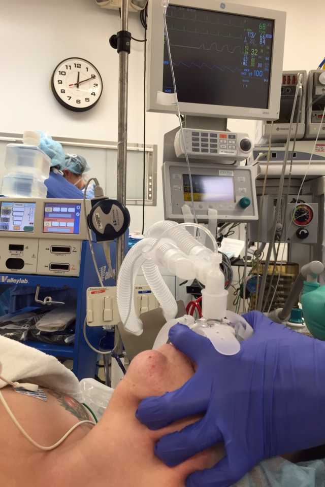
Photo: GlideScope intubation with Nasal TsePAP mask in place  
Courtesy of Dr. James Tse


II   High Flow Nasal Oxygen

Anil Patel and S Nouraei  have shown (1.) that apneic patients can be maintained for extended periods of time up to 16 minutes by use of Very high flows of nasal humidified Oxygen. Obesity significantly reduced the effectiveness to the 5-7 min range of time above 90 O2 SAT in some patients.
Patients were 40 degrees head up and had 70 L per min. of humidified O2 delivered through the Optiflow delivery system.

1. Anaesthesia 2015, 70, 323–329

Tuesday, June 30, 2015

Conventional Versus Video Laryngoscopy for Tracheal Tube Exchange. Glottic Visualization, Success Rates, Complications, and Rescue Alternatives in the High Risk Difficult Airway Patient.

Tom Mort has focussed on the advanced technique of using Video Laryngoscopy to monitor the high risk activity of exchange catheter use. The video images allow timely rotation or manipulation of the Tube and AEC to assure completion of the procedure.

Mort, Thomas C. MD , Braffett , Barbara H. PhD.
Hartford Hospital , Hartford Connecticut.

    Building on previous work showing the increased level of complications associated with multiple attempts at intubation and demonstration that video laryngoscopy could be used effectively in conjunction with an exchange catheter ( AEC ) for endotracheal tube exchange, this work consolidates earlier ideas. (1-2).

Abstract:
BACKGROUND: Tracheal tube exchange is a simple concept but not a simple procedure because hypoxemia, esophageal intubation, and loss of airway may occur with life-threatening ramifications. Combining laryngoscopy with an airway exchange catheter (AEC) may lessen the exchange risk. Laryngoscopy is useful for a pre-exchange examination and to open a pathway for endotracheal tube (ETT) passage. Direct laryngoscopy (DL) is hampered by a restricted "line of sight"; thus, airway assessment and exchange may proceed blindly and contribute to difficulty and complications. We hypothesized that video laryngoscopy (VL), when compared with DL, will improve glottic viewing for airway assessment, and the VL-AEC method of ETT exchange will result in a reduction in airway and hemodynamic complications in high-risk patients when compared with a historical group of patients who underwent DL + AEC-assisted exchange.
METHODS: Critically ill patients requiring an ETT exchange underwent DL-assisted pre-exchange airway assessment. If the DL-assisted pre-exchange assessment rendered a "poor view," these patients underwent a VL-based airway assessment followed by a VL-assisted ETT exchange procedure. The DL and VL pre-exchange assessments were compared. The attempts, complications, and rescue devices required for ETT exchange were analyzed. These exchange results were then compared with a historical control group of patients who (1) were classified as a poor view on DL-assisted pre-exchange airway assessment; and (2) underwent a DL + AEC-assisted exchange. The airway assessment and ETT exchange were performed by a board-certified anesthesiologist from the Department of Anesthesiology alone or with anesthesia resident assistance.
RESULTS: Three hundred twenty-eight patients with a poor view on initial DL examination underwent a subsequent VL with comparison of views with the 337 patients in the historical control group (DL + AEC). A majority (88%) had a "full or near-full view" on VL examination. The first-pass success rate for ETT exchange was greater in the VL group (91.5% vs 67.7% with DL; P = 0.0001) and the number of patients requiring 3+ attempts was lower (1.2% vs 6.8% with DL; P = 0.0003). A commensurate difference in the incidence of mild and severe hypoxemia, esophageal intubation, bradycardia, and the need for rescue airway device intervention was also observed with VL exchange procedures when compared with the historical DL + AEC group.
CONCLUSIONS: These findings support the hypothesis that VL may result in better glottic viewing for airway assessment and may permit the ETT exchange procedure to be performed with fewer airway and hemodynamic complications. Execution of the ETT exchange over an AEC was augmented by improved glottic visualization to allow more efficient and timely ETT passage. Multiple attempts to resecure the airway increased the number of exchange complications. VL + AEC exchange led to fewer attempts and is consistent with the recommendation of the American Society of Anesthesiologists Difficult Airway Task Force to limit laryngoscopic attempts and, as a consequence, decrease complications. A VL-based pre-exchange airway assessment may be a valuable procedure for both planning the exchange and uncovering unrecognized airway maladies, for example, partial or complete self-extubation.

References:

1.Emergency Tracheal Intubation: Complications Associated
with Repeated Laryngoscopic Attempts
Thomas C. Mort, MDDepartment of Anesthesiology, Hartford Hospital, University of Connecticut School of Medicine
Anesth Analg 2004;99:607–13


2.Tracheal tube exchange: feasibility of continuous glottic viewing with advanced laryngoscopy assistance.Mort TC. Anesth Analg. 2009 Apr;108(4):1228-31. doi: 10.1213/ane.0b013e3181990a82.

Thursday, June 4, 2015

A randomized multi-institutional crossover comparison of the GlideScope® Cobalt Video laryngoscope to the flexible fiberoptic bronchoscope in a Pierre Robin manikin

  1. Pediatric Anesthesia   
  2. John E. Fiadjoe1,*, 
  3. Matthew Hirschfeld1, 
  4. Stephan Wu1, 
  5. James Markley1, 
  6. Harshad Gurnaney1, 
  7. Abbas F. Jawad1, 
  8. Paul Stricker1, 
  9. Todd Kilbaugh1, 
  10. Patrick Ross2 and
  11. Pete Kovatsis3

Article first published online: 27 APR 2015
DOI: 10.1111/pan.12668

The Manikin study reported by John Fiadjoe et. al. illustrates findings that possibly indicate ease of learning the device handling features of equipment rather than effectiveness of the VL and FOB devices in the Pierre Roban complex . The jump from Manikin to Actual patient care is significant and is affected by secretions, relaxation, and muscular tone. 
This study did show that an institutional difference in results was apparent suggesting the need for disciplined training to achieve optimal results. The people are being measured as well as the devices in question. This study comes from a very experienced group of paediatric airway managers.

Summary

Background

The GlideScope Cobalt Video laryngoscope is being used more often in children with challenging laryngoscopy. There are, however, no pediatric trials comparing it to flexible fiberoptic bronchoscopy, the current accepted gold standard. This preliminary manikin study compares the first-attempt intubation success of the GlideScope Cobalt video laryngoscope to the flexible fiberoptic bronchoscope when performed by attending pediatric anesthesiologists at two major pediatric centers.

Methods

This prospective randomized, crossover study evaluated 120 attempts (60 with each study device) to intubate the AirSim Pierre Robin manikin (PRM) with fiberoptic bronchoscopy and video laryngoscopy (VL). Attending pediatric anesthesiologists from two quaternary pediatric centers were eligible to participate. Each attending anesthesiologist randomly performed a single tracheal intubation attempt with one of the study devices followed by the alternate method. The primary outcome was the first-attempt success rate of tracheal intubation. Blinding was not feasible. We hypothesized that first-attempt success would be higher with fiberoptic bronchoscopy.

Results

Thirty anesthesiologists from each center were randomized to use one of the study devices followed by the alternate method. We analyzed all participants' data. There was no overall difference in first-attempt success between VL and fiberoptic bronchoscopy (88.3% vs 85% respectively, P = 0.59). There were significant institutional differences in first-attempt success using VL (76.7% vs 100%).

Conclusions

There was no difference in first-attempt success of tracheal intubation using VL vs fiberoptic bronchoscopy when performed by attending anesthesiologists at two large pediatric centers. However, institutional differences exist in success rates with VL across the two centers. Results from single-center device evaluations should be verified by multi-center evaluations. A significant proportion of attending anesthesiologists lack experience with advanced airway devices; targeted education may enhance intubation success and patient safety.

Wednesday, June 3, 2015

Tracheal intubation in patients with odentogenous abscesses and reduced mouth opening- REPEAT POST

British Journal of Anaesthesia 112 (2): 348–54 (2014)

M. Schumann1, I. Biesler1, A. Borgers1, R. Pfortner2, C. Mohr2 and H. Groeben1*
1 Department of Anaesthesiology, Critical Care Medicine and Pain Therapy and 2 Department of Oral and Cranio-Maxillofacial Surgery, Kliniken
Essen-Mitte, Henricistr. 92, Essen 45136, Germany.


* Corresponding author. E-mail: h.groeben@kliniken-essen-mitte.de


Background. Odentogenous abscesses with involvement of the facial or cervical spaces can be

life-threatening andoften have to be drained under general anaesthesia. Trismus andswelling

can make intubation with a Macintosh laryngoscope difficult or even impossible. However,
indirect laryngoscopy has been successful when conventional direct laryngoscopy has
failed. Therefore, we evaluated the efficacy of the Glidescope laryngoscope in patients with
odentogenous abscesses and the improvement in mouth opening after neuromuscular block.
Methods. After approval of the ethics committee, 100 patients with odentogenous abscesses
were randomized to undergo tracheal intubation with the Glidescope or Macintosh
laryngoscope. Success rate, visualization of the glottis, intubation duration, and need for
supporting manoeuvres were evaluated.
Results. Intubation with the Glidescope was always successful, while conventional intubation
failed in 17 out of 50 patients (P,0.0001). In all patients in whom conventional tracheal
intubation failed, a subsequent attempt with the Glidescope was successful. The view at the
glottis (according to Cormack and Lehane; P,0.0001), intubation duration [34 s (CI 27–41)
vs 67 s (CI 52–82), mean (95% confidence interval); P.0.0001], and need for supporting
manoeuvres (P,0.0001) were significantly different. The inter-incisor distance improved
overall with induction of anaesthesia from 2.0 cm (CI 1.8–2.2) to 2.6 cm (CI 2.3–2.9;
P,0.0001) and was correlated with the duration of symptoms.
Conclusions. In patients with odentogenous abscesses, the use of a Glidescope laryngoscope
was associated with significantly faster tracheal intubation, with a better view, fewer
supporting manoeuvres, and a higher success rate than with a conventional laryngoscope.
Improvement of the inter-incisor distance after induction of anaesthesia correlated with
the duration of symptoms.
Keywords: intubation, difficult; intubation, tracheal; laryngoscope, Glidescope; laryngoscope,
Macintosh
Accepted for publication: 26 June 2013